The transmission electron microscope is a laboratory equipment, able to transmit and diffuse from electrons to form images, so that in this way, information about the crystalline structure can be reached, through the emission of X-rays, to know the elemental structure of the sample.
Similarly, with this instrument, it allows to obtain increments of 1000 000x to appreciate from chromosomes and DNA molecules (deoxyribonucleic acid), to atoms with a resolution power of 0.2 nm.
In this sense, the adaptation of this new equipment allows us to observe biological samples, such as the ultramicrotome and the diamond blade, allowing us to obtain ultra thin tissue cuts that resist the electron beam and high vacuum, for histological procedures with chemical fixators in the samples.
Transmission Electron Microscope Origin
It’s been more than 340 years since Antony Van Leeuwenhoek, with the invention of his simple microscope, discovered sperm cells in 1677, years in which the lens system of a microscope made up of optical light or photons has been perfected, to reach microscopes as sophisticated as phase contrast or fluorescence; but in 40 years or more, scientists perfected the transmission electron microscope, at that time, the world of biological sciences expanded with the invention of electron microscopy, achieving to observe images inside a cell, or particles of virus amplified thousands of times.
Currently, the team has become a fundamental tool in scientific research, in different areas of biology, medicine, materials and nanoparticles.
Transmission Electron Microscope Features
It is an instrument used for scientific research, weighing about a ton, the column around is 1.5 meters high, where high voltage is used to cause and guide a beam of electrons accelerated in high vacuum, which upon impacting one of the faces of the sample ultra thin tissue, form an image when emerging from the opposite face.
It is composed of the column, which transmits the electron beam, a high vacuum system, a cooling system, power currents and an image recording system, which is projected in two lengths on a fluorescent screen, and can be finally obtained through a photographic film or a digital camera in a computer.
Practices using the Transmission Microscope
Transmission electron microscopy can be used for the determination of microbial diseases, although its main application is in research. It provides ultra structural details, advantageous for the identification of viruses, bacteria, parasites and fungi. The greatest involvement is in the rapid analysis of viral diseases and different types of clinical samples can be explored, such as blood, feces, urine, biopsies, body fluids, etc.
The applications of the technique are numerous in both material science and biomedical science, which are:
- Determination of the morphology, shape, dimensions and position of microcrystals or particles observed in the sample.
- The crystallography is due to the position of the crystalline planes, study of defects, etc.; as well as the chemical composition of the material.
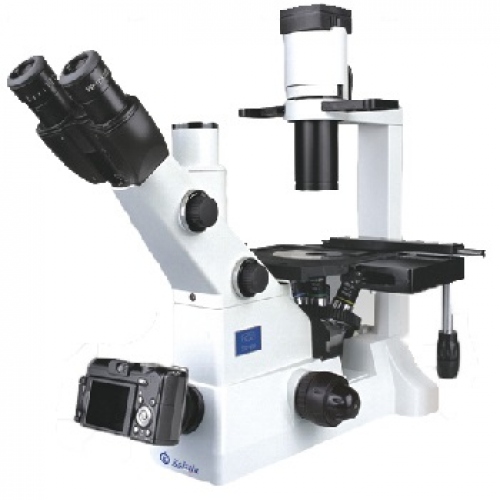
Kalstein brand microscope
At Kalstein, we have equipment suitable for laboratories, and in compliance with our users, we offer you the Microscopes, belonging to the YR models, with very attractive general characteristics, such as; fly-eye lens lighting. Infinite optical system. Tilt binocular display head. Fivefold backward (no coding). Infinity siedentopf binocular display head; inclined to 45° interpupillary 47-78 mm. Infinite phase contrast lens. Inserted Abbe capacitor NA1.25 (including empty plate). 3 W S-LED (LCD screen amplification), sleep timer, brightness lock and indication, etc., Two wave ranges (B, G, U, V can be combined). application software. HERE
We are manufacturers and we have the best advice, so that your purchase is the ideal and at excellent prices. For more information, visit our website HERE