The Human Papillomavirus (HPV), belonging to the family papovaviridae, is a relatively small intranuclear replication virus with a diameter of approximately 50 nm. HPV is a sexually transmitted infection that occurs frequently in most countries of the world, is detected 30 to 40% of cases in women of childbearing age (symptomatic and asymptomatic), and is associated with pre-neoplastic disorders and invasive carcinoma of the cervix. A family of more than 200 papillomavirus has been identified, which can infect mucous membranes and skin in almost all vertebrate species. Sometimes these lesions are not visible and are detected only through special examinations.
Importance of the HPV test
HPV is able to affect the host cell by the reproduction process so that it transforms into a malignant cell, and becomes a cancer cell. There are many cases in which women who are tested negative for cervical cancer usually also have human papillomavirus infections. Early identification of the virus is one aspect that guarantees successful treatment. In addition, regular gynecological check-ups are indispensable to prevent the progressive development of the virus, because it can go a long time without being detected, causing infections to be persistent and can become fatal.
Using a Microscope to Detect the Virus
It was in 1950 that HPV was first observed in skin papilloma samples. But it was until 1983 that the researcher Harald Zur Hausen replicated the outer layer of the virus, and managed to isolate under the microscope two strains responsible for 70% of cancers in the cervix. Their work contributed greatly to the development of the vaccine that prevents HPV-related infections. Historically, the use of microscopes in medical science has led to a breakthrough in understanding and understanding the virus, and continues to be a preferred method of tracking. Nowadays genetic engineering under the microscope studies how it relates to the oncogenic potential of the virus for the development of antiviral drugs.
Microscopic techniques are a useful tool that can be used to determine papillomavirus particles in samples of scrapings of exophytic warts from biopsies performed by the specialist. Once the virus has been detected, it is extremely important to know how it works, its morphology, which genomic family it belongs to and how advanced it is.
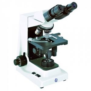
What is the microscope? And what is its function?
The microscope is an optical instrument that, thanks to its resolution power, has the ability to enlarge images of objects that are not visible to the human eye.
There are a variety and types of equipment for microscopy. These instruments are very useful in the field of scientific research. In the particular case of medicine, biology and clinical laboratories allow the study of microscopic structures of living and non-living matter. They are tools that make it possible to observe the morphology of cells, viruses, bacteria, and small organisms such as larvae, mosquitoes and their eggs, among others.
Types of optical microscopes
There are several types of microscope designs:
- Stereoscopes or magnifying glasses: they have the unique ability to see objects in three-dimensional, usually with employees in laboratories, they can see grains, small rocks, small organisms, etc.
- Optical: it is used to observe living organisms or objects not visible to the naked eye, it allows to see more broadly the smallest thing, such as multicellular cells, protozoa, etc.
- Electronic: Uses an electron beam instead of a light beam to form an image. It has a great depth of field, which allows you to focus at the same time a great detail samples at micrometric scales and manometer. They are very useful for studying organisms much smaller than with optical microscopes can not be observed. HERE
At KALSTEIN we are MANUFACTURERS, we offer you a variety of types of optical microscopes, with cutting edge technology. If you need to know more about our equipment, about PRICE, BUY or SELL, visit us at HERE