It is a progressive disease related to cognitive impairment, memory loss, and other important mental functions. It occurs because the connections of brain cells and the cells themselves degenerate and die. Currently, no cure has been found for this disease, but early detection contributes to an effective control treatment. Alzheimer’s represents about 80% of dementia cases diagnosed in elderly people, is recognized as a neurodegenerative disorder more common in old age, and one of the most common causes of dementia in the world. According to special microscopy studies based on the morphology and monitoring of structural development, they have determined that naturally occurring proteins fold incorrectly and are grouped into smaller filaments and groups known as oligomers. Scientific research has found that these structures are toxic and that they impair or eliminate neurons.
The microscope and Alzheimer’s disease
The first histology of this disease was made by Lois Alzheimer on November 4, 1906, who presented the microscopic study of the cerebral cortex of a patient who died while being treated for a type of cenile dementia at the XXXVII Conference of Psychiatry of the German Southwest in Tübingen. Later this disease would be known as Alzheimer’s, which is directly related to changes in the structure of the cerebral cortex, being possible to observe through the microscope:
- Brain tissue has far fewer neurons and synapses than a healthy brain.
- Presence of fibrillary lesions called neuritic plaques. Abnormal groups of protein fragments accumulate between neurons forming amyloid plaques.
- tangles of damaged and dead neurons, composed of small intertwined and twisted fibrils within neurons of another protein, located mainly in neocortex and hippocampus, also called neurofibrillary tangles and cortical atrophy.
Histologic techniques and observation under a microscope: staining
Most cellular tissues are colorless to the lens, which is why it is necessary to stain them to observe their morphological characteristics with the optical microscope. Histologic techniques are a series of steps to prepare the tissue for viewing under a microscope. The staining is one of the steps to perform. This is achieved by applying on tissue sections dyes, substances capable of binding to tissue structures providing color. The molecule of a dye usually has two main components: the substance that provides the color, called chromogen, and another that allows binding to tissue called auxochrome.
Some staining techniques used to determine histopathologic lesions in the brain using a microscope
Commonly used staining techniques to evidence histopathological lesions in the brain with Alzheimer’s disease in paraformaldehyde-fixed tissue:
- Silver staining: This technique uses the affinity of silver for the β-folded conformation present in the deposits of Αβ fibrilar and neurofibrilar tangles to identify lesions.
- Congo Red Staining: This is a direct staining with different affinities for fibrillary and non-fibrillary materials. It has the disadvantage that it can be difficult to visualize if the sample has a low sensitivity.
- Thioflavin-S: Considered an advantageous and simple technique because they can mark neuritic plaques and neurofibrillary tangles.
- Immunohistochemical staining: In this technique, it is essential as a diagnostic tool, using specific antibodies to observe the amount, distribution in the tissue and cellular location of immunogenic epitopes in sections of tissue fixed in formalin.
- Thiazine Red: Its functional feature is to bind to fibrillary forms with folded beta conformation. It consists of a staining made with the red dye thiazine, which is a naphthol-derived structure, produces an excitation fluorescence over a range of 530-560 nm. HERE
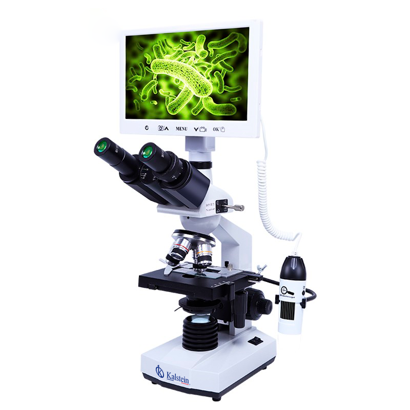
Microscopy is a technique that offers wide advantages, but its scope in research depends on the experience of the analyst and the equipment used. At KALSTEIN we are MANUFACTURERS, we offer you variety in types of optical microscopes, with cutting edge technology. If you need to know more about our equipment, about PRICE, BUY or SELL, visit us at HERE

