In histology laboratories some staining techniques are used to detect possible pathogens or cellular components that will help pathologists to determine the existence of diseases, there are a significant number of special stains of common application, with reagents and prepared solutions, many of these special stains have applications for microwaves, so you can get the results in a fraction of the time.
Histology being the study of tissues, requires great attention in its microscopic structure, development and functions, for this there are a number of techniques that facilitate the study of structures not visible to the human eye and staining is one of them; facilitating the observation of the different structures and substances of the tissues, so an automatic tissue dyeing equipment is indispensable in a clinical laboratory of pathology.
Tissue staining in histology
Although the observation is not considered as steps of the histological techniques but as the final objective of them, since the function of histology is to observe the lamellae under a microscope to obtain a diagnosis, and in summary what is used to arrive at that diagnosis, this is the case of tissue staining, since the cells, by themselves, do not have coloration; here highlights the importance of staining, since it allows us to observe the tissue morphology.
Although there are many types of stains that help differentiate the structure or substances of tissues, one of the most commonly used is routine staining or hematoxylin and eosin (H&E), which uses a dye called hematoxylin that stains acidic substances or substances that contain them, such as the nucleus containing deoxyribonucleic acid (DNA) and yellowish eosin stains basic structures such as the cytoplasm and other eosinophilic organelles of the cell.
In vivo and in vitro staining
An in vivo staining or vital staining, in this case it is a process that is performed to stain living tissues, the vital staining has as purpose to reveal the cyto-structure and is able to determine the presence of chemicals within the cells or tissues, this staining with contrast causes some cells or structures to acquire colors to then be able to study their location and morphology while they are in operation.
An in vitro staining or “supravital” this is the case where the coloring cells have been removed from their biological context and uses a combination of several colorants to achieve more details and characteristics, this technique combined with specific preparation methods of sample fixation, works as a repeatable and consistent diagnostic tool.
In vitro staining methods
The preparation methods used in a stain also depend on the analysis you want to perform, however, there are some stages that could be present in some or almost all:
Fixation: consists of modifying the physicochemical properties of proteins that are present in a cell or tissue, and at the same time preserving as much as possible the forms of that cell or tissue, another use of this technique is to kill, adhere and alter by means of heat a specimen so that it accepts staining; for this process a chemical fixative is used.
Inclusion: process where the tissues can be embedded in paraffin or some polymer in order to increase their strength and stability, also allows to cut the small blocks of tissues in extremely thin slices and thus obtain more definition in the microscopic images.
Permeability: In this process, cells are treated with a mild surfactant, in order to dissolve the cell membrane to allow large dye molecules to access the structures inside the cells.
Assembly: is the process where histological sections adhere to the glass slide and has the purpose of increasing the mechanical strength of the sample, and in this way can perform the observation or analysis in the microscope.
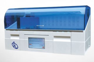
KALSTEIN brand automatic fabric dyeing
The intelligent automatic water inlet/outlet/outlet control system ensures sufficient washing performance at each step and improves water efficiency.
High quality imported parts, smooth operation, low noise, ergonomic design
Fully intelligent design, enabling timely determination and automatic recovery of an abnormal event
The wide LCD touch screen and convenient human machine interfaces provide the user with clear and sufficient information on the working status (Online Help)
Processing time is automatically calculated and displayed on the screen, allowing the user to create a more efficient workplan
4 sets of editable Chinese and English programs can be stored on the system and viewed online
Intelligent automatic water inlet/outlet/outlet control system ensures sufficient washing performance at every step and improves water efficiency
The 36 processing and staining protocols can be programmed and stored in the system, including the operation error alert function
Green inner cycle air purification system to absorb and remove poisonous gas
Visual simulation in real time with icons that show the working state dynamically, in a clear and intuitive way.
If you want to buy any of the automatic tissue staining equipment for histology laboratories, please be aware to contact the best ones, this service we can offer you through our online channels and at the best PRICE in the market, also if you want to know the catalog of high-end products that KALSTEIN have for you visit us HERE we assure you that through our online PURCHASE channels that are very easy and viable from anywhere in the world, reminding them that we are a manufacturer of high-level laboratory equipment for sale. HERE