Hepatitis is a disease characterized by affecting the liver, causing inflammation. It can be acute or chronic type, if the inflammation is recent is said to be acute hepatitis, however, if inflammatory processes last more than six months, then it is spoken of chronic hepatitis. All hepatitis is communicable disease, which means it can be prevented. For example, hepatitis A and E viruses are spread by contaminated food and water, so good hygiene and clean water are the ways to prevent them. On the other hand, hepatitis B and C are transmitted through sexual intercourse or blood contact.
The hepatitis virus is detected by simple blood tests that look for antibodies. When a person is infected by coming into contact with one of the viruses that cause hepatitis, the immune system produces antibodies, so if those antibodies are present in the blood at the time the blood test is being done, it means that the person was in contact with the virus at some point in his or her life, or that he or she is infected. On the other hand, the way in which these viruses were described and characterized was through the use of the electron microscope.
Electron microscope
The electron microscope is a large piece of equipment whose foundation is based on the use of an electron beam to illuminate the sample. The wavelength of electrons is much shorter than that of visible light, which is why the electron microscope can show smaller structures and agents, such as viruses, that are impossible to see with a conventional microscope. A conventional microscope works with visible light, whose shortest wavelength is about 4,000 angstroms, unlike the wavelength of electron microscopes, which is about 0.5 angstroms.
The electron microscopes have several components, have a cannon electrons through which they are emitted to collide with the sample and thus create a magnified image. In addition, they have magnetic lenses that create fields with which they focus and direct electrons. On the other hand, they have a vacuum system so that the air molecules do not deflect electrons, which is why this is an indispensable piece. Finally, all electron microscopes have a recording system of the image produced by electrons.
Use of a microscope to screen for hepatitis B virus
Hepatitis B is a primarily sexually transmitted liver disease, with an estimated 296 million people suffering from the disease in 2019, resulting in 1.5 million new infections annually. In 2019, the virus killed 820,000 people. For this reason it is necessary the continuous investigation of these viruses and for them, the electron microscopy has become a powerful ally in the laboratories of virology and clinical pathology.
Electron microscopes are indispensable equipment in the description of any virus and hepatitis B virus is not exempt from this. Hepatitis B virus (HBV), an agent of the family of Hepadnaviridae, the cause of the disease known as hepatitis B, is a small virus, measures approximately 42 nm, so it is impossible to appreciate it with a conventional microscope, since it has a resolving power of 0.2 µm, while the resolving power of optical microscopes is far below this. Thanks to the invention of the electron microscope, hepatitis viruses, including the hepatitis B virus, which was discovered by Dr. Baruch Samuel Blumberg in 1963, could be photographed and described.
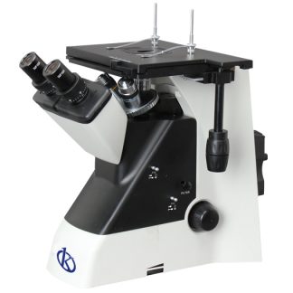
Kalstein-branded microscopes
Kalstein is a leading company dedicated to the sale of clinical and laboratory equipment, however, we are also characterized by being MANUFACTURERS of our products, so we assure you that they are of excellent quality and high technology, of course, at the best PRICES in the market. We have a wide range of microscopes, from stereoscopic models to biological, metallurgical and inverted microscopes. To make the PURCHASE of one of our microscopes, just enter our catalog: HERE

