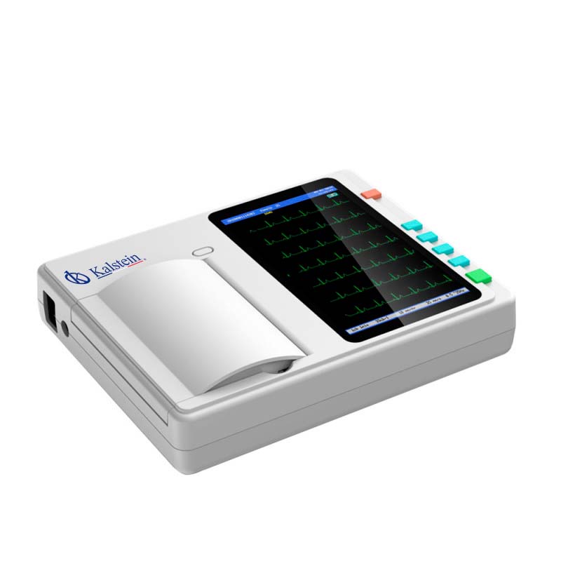
It is a medical device, charged with recording the negative and positive cardiac activities of patients, driven by electrodes, able to record the damage of the heart, the speed of palpitations or some anomaly, in addition to the effects left by the cardiac drugs.
This type of device is characterized by having 3.6 or 12 channels and recording each of the 12 leads in a period of 2.5 seconds. In addition, it offers many advantages over single-channel tests, as they support cross-checking multiple bypass examinations during the same heartbeat, providing much interpretation, and optimizing the accuracy of the diagnosis accordingly.
Functions of a Multichannel Electrocardiograph
This equipment allows to recognize and differentiate by means of a computer with patterns, the normal ECG signals from those that are not. It determines and identifies important measurements, by analyzing heart rate, signal amplitude, wave capacity, or the intervals between the wave mechanisms.
It also has a connector of deviations, where the pattern certifies the contribution of each electrode through resistances, achieving the derivation of interest. By using these equipment, and a memory system, signals are stored next to the data entered manually, and by means of a digital converter, the printed reading is achieved. Subsequently, it has a micro controller, which inspects all registered electrocardiographic devices.
Parts of an electrocardiograph
This team presents the mechanisms to perform the electrocardiograms of patients, conformed as follows:
- It has an amplifier of biopotentials, which emits signals of calibration, protection, preamplifier, isolation circuit and amplifier manager.
- It reduces the impedance of the ground electrode, by connecting the right leg, creating an active earth removed from the electric ground, in order to increase safety.
- Shunt selector, through a is an amplifier pattern of biopotentials, accessing the transmission of the electrodes to achieve the derivation of interest.
- It has a storage system, which reserves the signal before it is printed.
- Microcontroller: to record the operations performed by the electrocardiograph.
- It must be operated by a specialist, who is responsible for printing and reading the signals captured, also using pens and continuous thermal paper or inkjet.
According to the above, each circuit must be enforced to obtain the desired results, which are captured by V6 unipolar bypass with the electrodes located on the top two extremities and on the left foot, which are sent to the microcontroller. Subsequently, the original signal is passed through to properly prepare the ECG socket for a better analysis, which applies the 80 Hz filter, and eliminates any magnetic interference that may be present in the environment. Also, thanks to a 20 Hz filter, it sends important information and eliminates 5 Hz signals, similar to those emitted by the lungs. Finally, a filter between 55 and 65 Hz is applied, which removes external frequencies such as that generated by the appliance’s power supply.
Electrocardiograph brand KALSTEIN
We at KALSTEIN have equipment trained to perform electrocardiograms, commonly used to detect abnormal heart rhythms and investigate the causes of chest pains. An electrocardiogram also records the electrical activity of the heart. The heart produces tiny electrical impulses that spread through the heart muscle to cause the heart to contract. Therefore, we offer you 12-channel ECG Electrocardiographs, where you can simultaneously collect 12-lead ECG signals and print ECG waveforms with a thermal printing system. HERE
We have YR models, with 7″ touch screen, supports Ecg analysis capabilities, 12 standard conductors, input circuit current ≤0.0.05ì, anti-baseline drift automatic, 12.5 mm/s, 25 mm/s, 50 mm/s. For more information, visit us at HERE


