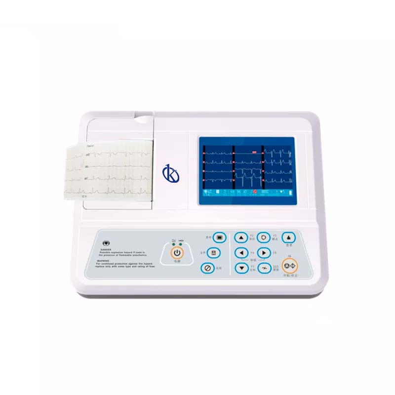
An electrocardiograph of this type is characterized by having 12 channels and recording each of the 12 leads within 2.5 seconds; they allow the visualization of the analysis in real time before printing the records, providing efficiency and improving the accuracy of the patient’s diagnosis; In this way you avoid having to repeat the procedure, a 12-lead electrocardiograph has advantages over other types, such as the ability to send data to the ECG administration system without printing.
This 12-lead device helps diagnose abnormalities in electrical cardiac conduction, through the electrodes that transfer the impulses to an ECG monitor, the 12 leads provide waves from 12 different angles or views of the heart in an instant, one electrode is placed on each of the four extremities and another six in specific places of the chest, making a total of 10 electrodes in the patient’s skin.
12 leads
Derivations means electrodes that record the electrical activity produced by the cells and the electrocardiograph converts them into waves, thus analyzing a certain part of the heart and thus establishing whether that part has suffered an injury.
The 12 derivations are:
Bipolar derivatives members I, II, III:
- Bypass I: It records the voltage difference between the left arm and right arm electrodes.
- Bypass II: The voltage difference between the electrodes of the left leg and the right arm.
- Bypass III: The voltage difference between the left leg and left arm electrodes.
Increased derivations of members:
- aVR placed on right arm
- aVL placed on left arm
- aVF placed on left foot
The precordial leads V 1 to V 6 are placed in such a way that the record of electrical activity can divide the heart into a horizontal plane.
How to detect an abnormality in the heart
It is important to know how to read the record of these derivations, which provides this electrical record correlates with the structures of the heart so that alterations in any of them give us an idea of the location of the causal defect; through them we can detect where exactly the problem or anomaly is located, we have the following:
- V1-V2 Septal wall
- V3-V4 Previous Wall
- I, aVL ,V5-V6 Side wall
- II, III, aVF Bottom wall
How does it work?
In addition to indications of how to place an electrocardiogram, its operation is really simple, electrocardiography records and measures electrical impulses that occur during the contraction of the heart muscle that are generated by the heart.
The operation of the electrodes is to capture the electrical activity that occurs in the tissues below where they are placed in the body part, in a general way they function as vectors, that is, as a whole they measure the amount of energy and the direction of an electrical impulse, when an impulse approaches the electrode, it is recorded as a positive wave, on the contrary, it happens when the impulse moves away from the electrode because then the record will be of a negative wave.
Characteristics of the 12-lead electrocardiograph kalstein mark
The 12-channel ECG machine, branded KALSTEIN is an electrocardiograph that can collect 12-lead ECG signals simultaneously and print ECG waveforms with a thermal printing system. The electrocardiogram is commonly used to detect abnormal heart rhythms and to investigate the cause of chest pains. An electrocardiogram records the electrical activity of the heart. The heart produces tiny electrical impulses that spread through the heart muscle to cause the heart to contract. These impulses can be detected by the ECG machine.
If you want to know the catalog of high-end products that we KALSTEIN have for you, visit us HERE we have a specialized team that will answer your doubts, remember that choosing the best and most updated Electrocardiograph of 12 leads, guarantees the health center to offer the patient a safe and reliable examination, in addition we assure that through our online sales channels it is very easy and viable to get the best prices of the market, remember we are Company manufacturer of Laboratory Equipment recognized worldwide. HERE


